Lysosome Structure and Function

Lysosome drawing easy colored – Lysosomes are membrane-bound organelles found in animal cells, crucial for maintaining cellular health through their role in waste recycling and degradation. Their structure and function are intricately linked, enabling them to perform their vital tasks within the cell.
Lysosome Structure
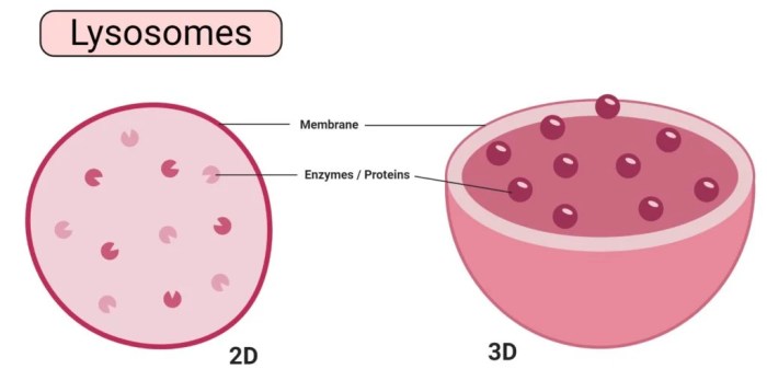
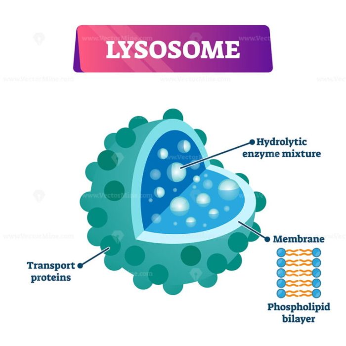
Lysosomes are spherical organelles enclosed by a single lipid bilayer membrane. This membrane is vital for maintaining the acidic pH (around 4.5-5.0) within the lysosome, which is essential for the activity of the hydrolytic enzymes they contain. The interior of the lysosome is filled with a variety of hydrolytic enzymes, including proteases, nucleases, lipases, glycosidases, and phosphatases. These enzymes are capable of breaking down various biological macromolecules, such as proteins, nucleic acids, lipids, and carbohydrates.
The lysosomal membrane also contains specialized proteins that help transport the breakdown products out of the lysosome and protect the lysosome itself from self-digestion.
Lysosome Function in Cellular Digestion and Waste Removal, Lysosome drawing easy colored
Lysosomes are the primary sites of intracellular digestion. They receive materials from various sources, including phagocytosis (engulfing extracellular materials), endocytosis (taking in substances from the cell’s environment), and autophagy (recycling cellular components). Once inside the lysosome, these materials are broken down by the hydrolytic enzymes into their constituent monomers, such as amino acids, nucleotides, fatty acids, and sugars.
These smaller molecules are then transported across the lysosomal membrane and reused by the cell in various metabolic processes. This process effectively removes cellular waste and recycles essential building blocks, maintaining cellular homeostasis.
Creating a simple, colored drawing of a lysosome can be a rewarding exercise in biological illustration. For a slightly different artistic challenge, consider exploring animal anatomy with a bat eared fox drawing easy tutorial; the techniques for depicting fur and form can be surprisingly transferable to the smooth, rounded shapes of a lysosome. Returning to our cellular subject, remember to highlight the lysosome’s membrane and internal enzymes for a truly informative depiction.
Autophagy Mediated by Lysosomes
Autophagy, meaning “self-eating,” is a crucial cellular process where damaged organelles, misfolded proteins, and other cellular debris are selectively targeted and degraded by lysosomes. The process begins with the formation of a double-membraned structure called an autophagosome, which engulfs the targeted material. The autophagosome then fuses with a lysosome, delivering its contents to the lysosomal hydrolytic enzymes for degradation.
This process is essential for removing potentially harmful cellular components and maintaining cellular health. Dysregulation of autophagy is implicated in various diseases, including cancer and neurodegenerative disorders. For example, impaired autophagy contributes to the accumulation of misfolded proteins in neurodegenerative diseases like Alzheimer’s and Parkinson’s.
Comparison of Lysosomes with Other Organelles Involved in Cellular Degradation
Lysosomes are distinct from other organelles involved in cellular degradation, such as proteasomes. Proteasomes are large protein complexes that degrade misfolded or damaged proteins primarily in the cytoplasm. Unlike lysosomes, proteasomes do not degrade lipids or carbohydrates. Peroxisomes, another type of organelle, are involved in the breakdown of fatty acids and detoxification of harmful substances through oxidative reactions.
While both peroxisomes and lysosomes contribute to cellular degradation, they have distinct substrate specificities and mechanisms of action.
Key Characteristics of Lysosomes
| Component | Function | Relationship to other organelles | Clinical Significance |
|---|---|---|---|
| Lipid bilayer membrane | Maintains acidic pH; regulates transport of enzymes and breakdown products | Receives materials from endosomes, autophagosomes, and phagosomes | Mutations affecting membrane proteins can lead to lysosomal storage disorders. |
| Hydrolytic enzymes (proteases, nucleases, lipases, etc.) | Degrade various biological macromolecules | Synthesized in the rough endoplasmic reticulum and processed in the Golgi apparatus | Deficiencies in specific enzymes can cause lysosomal storage disorders. |
| Acidic pH (around 4.5-5.0) | Optimal pH for hydrolytic enzyme activity | Maintained by proton pumps in the lysosomal membrane | Disruption of pH homeostasis can impair lysosomal function. |
| Transporters | Transport breakdown products out of the lysosome | Interact with other organelles to facilitate recycling of metabolites | Defects in transporters can contribute to lysosomal storage disorders. |
Simplified Lysosome Drawing Techniques

Creating a clear and informative drawing of a lysosome is crucial for understanding its function. This involves simplifying the complex internal structures while maintaining accuracy and visual appeal. By using basic shapes and strategic color choices, even a simplified drawing can effectively convey the key characteristics of this vital organelle.
Step-by-Step Guide to Drawing a Simplified Lysosome
Begin by drawing a slightly irregular circle or oval to represent the lysosome’s membrane. This shape reflects the lysosome’s dynamic nature and avoids a perfectly symmetrical representation which might be misleading. Next, add smaller, irregularly shaped circles and ovals within the larger shape to represent the hydrolytic enzymes contained within. These internal shapes should be smaller and more numerous to show the abundance of enzymes.
Finally, a few short, slightly curved lines can be added to the interior to represent the heterogeneous nature of the lysosomal contents. These lines should not be too prominent, lest they overwhelm the depiction of the enzymes.
Depicting the Lysosomal Membrane and Internal Contents
The lysosomal membrane can be represented with a thin, dark line surrounding the main oval. To emphasize its unique composition, a slightly textured line, or a line with small, subtle variations in thickness, could be employed. For the internal contents, avoid overcrowding. A few clearly depicted enzymes are more effective than a chaotic jumble. Varying the sizes and shapes of the internal circles and ovals suggests the diversity of the enzymes.
Consider adding a few small, dark dots to represent undigested materials within the lysosome.
Using Color to Represent Lysosome Components
Use a muted, light orange or yellow for the background to represent the cytoplasm surrounding the lysosome. The lysosomal membrane can be depicted in a dark brown or deep purple to highlight its crucial role in maintaining the integrity of the organelle. The hydrolytic enzymes within should be a different color, perhaps a lighter shade of purple or blue, to contrast with the membrane and the background.
Undigested materials can be represented with a darker shade of brown or black.
Creating a Visually Appealing and Informative Drawing
Maintain a balance between simplicity and detail. Avoid overwhelming the drawing with too much information. Use clear, concise labels to identify the key components: lysosomal membrane, hydrolytic enzymes, and undigested materials. The font size and style should complement the overall aesthetic of the drawing. Choose colors strategically to enhance clarity and avoid creating a visually cluttered image.
Three Variations of a Simple Lysosome Drawing
The following three variations showcase different aspects of lysosome function:
Variation 1 (Autophagy): This drawing depicts a lysosome engulfing a damaged organelle (represented by a smaller, faded circle). The damaged organelle is partially enclosed within the lysosome membrane, and the hydrolytic enzymes are shown more actively interacting with it. The overall color scheme is darker to reflect the breakdown process.
Variation 2 (Phagocytosis): This drawing shows a lysosome fusing with a phagosome (a vesicle containing a foreign particle, represented by a darker, irregularly shaped object). The foreign particle is being broken down within the lysosome, and the enzymes are depicted in a more concentrated manner near the particle. The colors used are brighter, reflecting the active engulfment process.
Variation 3 (Waste Breakdown): This drawing features a lysosome containing numerous small, dark circles representing various waste products. The enzymes are distributed throughout the lysosome, actively breaking down the waste materials. The colors are muted, reflecting the ongoing, less visually dynamic nature of waste processing.
FAQ: Lysosome Drawing Easy Colored
What are the best tools for creating a colored lysosome drawing?
Colored pencils, markers, or digital art software are all suitable. Choose the medium you are most comfortable with.
How can I add depth to my lysosome drawing?
Use shading and highlighting to create a three-dimensional effect. Consider overlapping shapes to suggest depth.
What are some common mistakes to avoid when drawing lysosomes?
Avoid overly complex details for introductory drawings. Focus on clearly representing the membrane and internal contents.
Are there online resources to help with lysosome drawing?
Yes, many online resources, including educational websites and scientific databases, offer images and diagrams of lysosomes.

