Understanding Animal Cell Structure
Animal cell coloring answer key biology corner – Animal cells, the fundamental building blocks of animals, are complex structures containing a variety of organelles, each with specialized functions crucial for cellular life. A thorough understanding of these organelles and their interactions is essential to comprehending the overall physiology of an organism.
Major Organelles and Their Functions
The intricate machinery of an animal cell relies on the coordinated action of numerous organelles. These structures work in concert to maintain cellular homeostasis, enabling the cell to carry out its vital processes.
| Organelle | Function | Description |
|---|---|---|
| Nucleus | Houses genetic material (DNA), controls cell activities. | The control center of the cell, containing chromosomes and the nucleolus, which is responsible for ribosome synthesis. |
| Ribosomes | Protein synthesis. | Small organelles found free in the cytoplasm or attached to the endoplasmic reticulum; they translate mRNA into proteins. |
| Endoplasmic Reticulum (ER) | Protein and lipid synthesis, detoxification. | A network of membranes; the rough ER (with ribosomes) synthesizes proteins, while the smooth ER synthesizes lipids and detoxifies substances. |
| Golgi Apparatus | Modifies, sorts, and packages proteins and lipids. | A stack of flattened sacs that processes and transports molecules synthesized by the ER. |
| Mitochondria | Cellular respiration, ATP production. | The “powerhouses” of the cell, generating energy through oxidative phosphorylation. |
| Lysosomes | Waste breakdown and recycling. | Membrane-bound sacs containing digestive enzymes that break down waste materials and cellular debris. |
| Vacuoles | Storage of water, nutrients, and waste. | Membrane-bound sacs that store various substances; generally smaller and more numerous in animal cells compared to plant cells. |
| Cytoskeleton | Maintains cell shape, facilitates movement. | A network of protein filaments (microtubules, microfilaments, intermediate filaments) providing structural support and enabling intracellular transport. |
| Centrioles | Organize microtubules during cell division. | Paired cylindrical structures involved in the formation of the mitotic spindle during cell division. |
The Cell Membrane and Its Role
The cell membrane, also known as the plasma membrane, is a selectively permeable barrier that encloses the cell’s contents. It regulates the passage of substances into and out of the cell, maintaining a stable internal environment. This membrane is composed primarily of a phospholipid bilayer, with embedded proteins that facilitate transport, cell signaling, and other functions. The fluid mosaic model describes the dynamic nature of this membrane, where components are constantly moving and interacting.
For example, receptor proteins on the membrane bind to specific signaling molecules, triggering intracellular responses. Ion channels selectively allow the passage of certain ions, contributing to membrane potential and cellular communication.
Organelle Interactions Within the Cell
Organelles rarely function in isolation; their activities are highly coordinated. For instance, proteins synthesized on the rough ER are transported to the Golgi apparatus for modification and packaging before being delivered to their final destinations. Mitochondria provide the ATP necessary for many cellular processes, including protein synthesis and transport within the cell. Lysosomes degrade worn-out organelles, recycling their components.
The cytoskeleton facilitates the movement of organelles and vesicles throughout the cell, ensuring efficient intracellular transport. The coordinated action of these organelles exemplifies the intricate organization and interdependence within the animal cell.
Need help with that tricky animal cell coloring answer key from Biology Corner? Sometimes, switching gears helps! Check out this awesome african animal coloring book for a fun break before tackling those organelles again. The vibrant colors might even spark some new ideas for your cell diagrams – you’ll be back to mastering those cell structures in no time!
Coloring Activities and their Educational Value: Animal Cell Coloring Answer Key Biology Corner

Coloring activities, often underestimated in higher education, offer a surprisingly effective pedagogical tool for reinforcing learning in cell biology. The tactile engagement and visual representation inherent in coloring significantly enhance comprehension and retention, particularly concerning complex structures like the animal cell. This method transforms passive learning into an active, multi-sensory experience.Coloring aids memorization and understanding of spatial relationships.
The act of carefully coloring each organelle within the cell’s boundaries fosters a deeper understanding of its three-dimensional structure and the relative positions of its components. This kinesthetic learning approach is particularly beneficial for visual learners, solidifying their understanding beyond rote memorization. Repeatedly tracing the Artikels and filling in the colors reinforces the shapes and locations of the organelles, leading to improved recall and spatial reasoning.
Effective Cell Coloring Worksheet Design
Effective cell coloring worksheets require careful consideration of several design elements. A well-designed worksheet should clearly illustrate the structure of an animal cell, accurately labeling each organelle and providing a color key. Overly complex designs, however, can be counterproductive, overwhelming the learner and hindering comprehension. Simpler, well-organized diagrams are generally more effective for learning. The choice of colors should also be considered; contrasting colors help distinguish different organelles and improve visual clarity.
For instance, using a bright green for the chloroplast (if applicable) and a deep blue for the nucleus aids in distinguishing them from other organelles. Worksheets that incorporate both labeling and coloring exercises provide a more comprehensive learning experience, combining visual and textual information to strengthen memory.
Learning Objectives Achievable Through Animal Cell Coloring
A well-designed animal cell coloring exercise can achieve several key learning objectives. The following points illustrate the potential educational value of such an activity:
- Students will be able to identify and label the major organelles of an animal cell.
- Students will be able to describe the function of each major organelle within the animal cell.
- Students will be able to accurately depict the spatial relationships between organelles within the animal cell.
- Students will demonstrate improved memorization and recall of animal cell structures and functions.
- Students will develop a deeper understanding of the overall structure and function of the animal cell.
Analyzing a “Biology Corner” Animal Cell Coloring Worksheet
A hypothetical “Biology Corner” animal cell coloring worksheet, while offering a potentially engaging introduction to cell biology, presents opportunities for improvement in terms of accuracy, clarity, and pedagogical effectiveness. Effective design requires careful consideration of common student misconceptions and potential sources of error. This analysis will identify strengths and weaknesses, suggest improvements, and address common student errors.Analyzing the Strengths and Weaknesses of a Sample WorksheetA well-designed worksheet should accurately depict the organelles of an animal cell, clearly labeling each structure and providing sufficient space for coloring and annotation.
Strengths might include a visually appealing layout, clear instructions, and a reasonable level of complexity appropriate for the target age group. Weaknesses, however, could include inaccurate representations of organelle size and shape, unclear labeling, or a lack of contextual information linking structure to function. For instance, a worksheet might incorrectly depict the Golgi apparatus as a simple sac rather than a complex network of flattened sacs, or it might fail to differentiate the rough endoplasmic reticulum from the smooth endoplasmic reticulum.
Ambiguous labeling can lead to confusion, especially for students who are new to the subject.
Potential Misconceptions Arising from Incorrectly Completed Worksheets
Incorrectly completed worksheets can reinforce several misconceptions. It’s crucial to address these to ensure accurate understanding.
The following points highlight common misconceptions that may arise from inaccurate worksheet completion:
- Oversimplification of Organelle Structure: Students might develop a simplified view of organelles, failing to appreciate their complex three-dimensional structures and internal organization. For example, believing the mitochondria is simply a bean shape, neglecting the cristae.
- Incorrect Spatial Relationships: The relative positions and sizes of organelles are crucial. An inaccurate depiction can lead students to believe organelles are randomly distributed or that their sizes are not proportionate. For instance, believing the nucleus is smaller than the cytoplasm.
- Confusion of Organelle Functions: Incorrect labeling or coloring can confuse the functions of different organelles. Students might incorrectly associate functions with the wrong organelle, leading to a flawed understanding of cellular processes.
- Misinterpretation of Cellular Processes: An inaccurate representation can impede understanding of how organelles interact and contribute to cellular processes like protein synthesis, energy production, or waste removal. This might lead to an inability to connect cellular structure to cellular function.
Correcting Common Errors in Student Worksheets
Effective feedback is crucial for correcting errors. Instead of simply marking answers wrong, instructors should guide students toward a deeper understanding.
Strategies for correcting common errors include:
- Providing Clear, Accurate Diagrams: Presenting students with accurate diagrams of animal cells, highlighting the correct structures and their relative sizes and positions, can correct misconceptions about organelle structure and spatial relationships.
- Encouraging Peer Review: Having students review each other’s work can help them identify and correct their own errors and learn from their peers’ mistakes.
- Using Interactive Models and Simulations: 3D models and simulations can enhance understanding of the three-dimensional nature of cells and organelles, addressing the oversimplification of structure.
- Connecting Structure to Function: Emphasis on the relationship between the structure of an organelle and its function can help students understand why certain structures are important.
Creating an Enhanced Animal Cell Coloring Worksheet
This section details the design of a superior animal cell coloring worksheet, addressing common shortcomings found in existing materials. The improved worksheet incorporates detailed descriptions of each organelle, clear instructions for completion, and a robust rubric for assessment, fostering a deeper understanding of animal cell structure and function.
The enhanced worksheet aims to move beyond simple coloring and engage students in a more active learning process. It achieves this through detailed descriptions that go beyond basic labels, promoting comprehension and retention of key concepts. The inclusion of specific instructions reduces ambiguity and ensures a consistent learning experience for all students. Finally, the rubric provides a clear framework for assessing student understanding, facilitating accurate evaluation of their learning outcomes.
Organelle Descriptions and Coloring Instructions
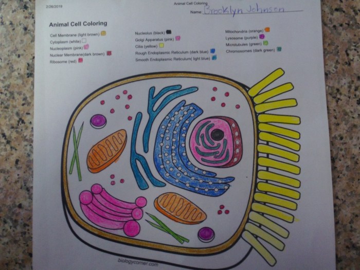
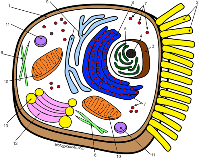
The worksheet features a large, clearly drawn diagram of an animal cell. Each organelle is numbered, and corresponding descriptions are provided in a separate section. These descriptions go beyond simple definitions, providing context about the organelle’s function within the cell and its interaction with other organelles. For instance, the description of the mitochondria would explain its role in cellular respiration and energy production, linking it to the cell’s overall metabolic activity.
Similarly, the Golgi apparatus description would detail its role in protein modification and packaging, connecting it to the endoplasmic reticulum. Coloring instructions specify a unique color for each organelle, promoting visual differentiation and aiding in memorization. Students are encouraged to use color-coding to highlight the functional relationships between organelles.
Student Instructions for Worksheet Completion
Students are instructed to carefully read the description for each numbered organelle before coloring it. They are encouraged to annotate the diagram with brief notes summarizing the organelle’s function. This active recall strengthens learning. For example, beside the colored mitochondria, a student might write “ATP production”. Further, students are asked to draw a simple flow chart illustrating the path of a protein from its synthesis in the ribosomes to its secretion from the cell, highlighting the involvement of the endoplasmic reticulum and Golgi apparatus.
This promotes a deeper understanding of the interconnectedness of cellular processes.
Rubric for Assessing Student Understanding
The rubric below provides a structured assessment of student work. It evaluates both the accuracy of coloring and the depth of understanding demonstrated through annotations and the flow chart.
| Criteria | Excellent | Good | Needs Improvement |
|---|---|---|---|
| Accuracy of Organelle Coloring | All organelles are correctly identified and colored according to instructions. | Most organelles are correctly identified and colored; minor inaccuracies present. | Significant inaccuracies in organelle identification and coloring. |
| Completeness of Annotations | Each organelle has a concise and accurate annotation describing its function. | Most organelles have annotations; some may be incomplete or slightly inaccurate. | Few or no annotations are provided, or annotations are significantly inaccurate. |
| Accuracy of Protein Pathway Flowchart | Flowchart accurately depicts the path of a protein from ribosome to secretion, including all relevant organelles. | Flowchart depicts the general path, but may have minor inaccuracies or omissions. | Flowchart is incomplete, inaccurate, or missing key elements. |
| Overall Neatness and Presentation | Worksheet is neatly colored and annotated, demonstrating care and attention to detail. | Worksheet is mostly neat; minor sloppiness may be present. | Worksheet is messy and disorganized, hindering understanding. |
Illustrative Examples of Animal Cell Structures

Animal cells, the fundamental units of animal life, exhibit a complex internal organization crucial for their diverse functions. Understanding the structure and function of key organelles is paramount to comprehending cellular processes. This section will delve into three critical organelles: the nucleus, mitochondria, and Golgi apparatus.
The Nucleus: Control Center of the Cell
The nucleus is the cell’s command center, housing the genetic material—DNA—and orchestrating cellular activities. Its structure comprises a double membrane, the nuclear envelope, punctuated by nuclear pores that regulate the passage of molecules between the nucleus and the cytoplasm. Within the nucleus, DNA is organized into chromosomes, condensed structures of DNA and proteins. A prominent structure within the nucleus is the nucleolus, the site of ribosome biogenesis.An accompanying image would show a large, roughly spherical structure (the nucleus) centrally located within the cell.
The nuclear envelope would be depicted as a double line, with small dots representing the nuclear pores. The nucleolus would appear as a denser, darker region within the nucleus. Chromatin (the less condensed form of DNA) would be represented as a diffuse, lighter-colored material filling the rest of the nuclear space. The visual representation would clearly illustrate the double membrane nature of the nuclear envelope, the selective permeability implied by the nuclear pores, and the distinct structure of the nucleolus, emphasizing the compartmentalization necessary for DNA replication and transcription.
The tightly packed chromosomes would be shown only in a cell undergoing mitosis, otherwise appearing as dispersed chromatin. This visual would underscore the nucleus’s role in safeguarding genetic material and regulating gene expression. The image would emphasize the importance of the nuclear envelope’s selective permeability in protecting the DNA and controlling the flow of information.The nucleus plays a critical role in DNA replication, the process of duplicating the genome before cell division, and transcription, the process of creating RNA molecules from DNA templates.
These processes are meticulously regulated within the nucleus, ensuring accurate genome duplication and precise control of gene expression.
Mitochondria: Powerhouses of the Cell
Mitochondria are the cell’s power generators, responsible for cellular respiration—the process of converting nutrients into energy in the form of ATP (adenosine triphosphate). Their structure is characterized by a double membrane: an outer membrane and a highly folded inner membrane called the cristae. The cristae significantly increase the surface area available for the electron transport chain, a crucial step in ATP production.
The space within the inner membrane is called the mitochondrial matrix, where the citric acid cycle takes place.An accompanying image would depict elongated, bean-shaped organelles scattered throughout the cytoplasm. The outer membrane would be a smooth, continuous line, while the inner membrane would be shown as a series of extensively folded, shelf-like structures (cristae) projecting into the mitochondrial matrix.
The matrix would be shown as a less dense region within the inner membrane, highlighting the compartmentalization of the processes involved in cellular respiration. The visual representation would emphasize the extensive surface area provided by the cristae, directly relating this structural feature to the function of ATP production. The different densities of the inner and outer membranes would also be visually distinct.
The image should clearly distinguish the outer and inner membranes, the cristae, and the matrix, emphasizing the spatial organization of the metabolic processes occurring within the mitochondrion.The intricate structure of the inner membrane, with its numerous folds, maximizes the efficiency of ATP production. The matrix contains enzymes essential for the citric acid cycle, further highlighting the compartmentalization of cellular respiration.
Golgi Apparatus: Protein Processing and Packaging Center, Animal cell coloring answer key biology corner
The Golgi apparatus, also known as the Golgi complex, is a series of flattened, membrane-bound sacs called cisternae. It acts as the cell’s processing and packaging center for proteins and lipids. Proteins synthesized in the endoplasmic reticulum (ER) are transported to the Golgi apparatus for modification, sorting, and packaging into vesicles for transport to their final destinations within or outside the cell.An image would depict a stack of flattened, membrane-bound sacs (cisternae) with associated vesicles.
The cisternae would be represented as parallel, slightly curved sacs, showing a clear delineation of their individual membranes. Smaller vesicles would be depicted budding from the edges of the cisternae, illustrating the process of protein packaging and transport. The image would emphasize the directional flow of materials through the Golgi apparatus, from the cis face (receiving end) to the trans face (shipping end).
The visual representation would clearly illustrate the progressive modification and sorting of proteins as they move through the Golgi stacks. The varying sizes and shapes of the vesicles would also be apparent.The Golgi apparatus’s layered structure facilitates the sequential modification of proteins as they move through the cisternae. This ordered process ensures that proteins are properly folded, modified, and targeted to their correct locations, emphasizing the importance of the Golgi apparatus in maintaining cellular organization and function.

