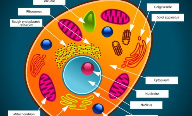
Animal Cell Structure & Function
Animal cell coloring questions key – Animal cells are the fundamental building blocks of animals, exhibiting a complex internal organization crucial for their survival and function. Understanding the structure and function of their organelles is key to comprehending how animals grow, reproduce, and maintain homeostasis.
Major Organelle Functions
The various organelles within an animal cell work together in a coordinated manner. Each organelle performs specific tasks essential for the cell’s overall operation. The nucleus houses the cell’s genetic material (DNA), controlling cellular activities. Mitochondria are the powerhouses, generating energy through cellular respiration. Ribosomes synthesize proteins, the workhorses of the cell.
The Golgi apparatus modifies, sorts, and packages proteins for secretion or use within the cell. The endoplasmic reticulum (ER) plays a crucial role in protein and lipid synthesis and transport. Lysosomes contain enzymes that break down waste materials. The cytoskeleton provides structural support and facilitates cell movement. The cell membrane regulates what enters and exits the cell.
Plant and Animal Cell Differences
While both plant and animal cells are eukaryotic (possessing a membrane-bound nucleus), they exhibit key differences. Plant cells possess a rigid cell wall providing structural support and protection, absent in animal cells. Chloroplasts, responsible for photosynthesis, are present in plant cells but absent in animal cells. Plant cells typically have a large central vacuole for storage, whereas animal cells have smaller, less prominent vacuoles.
These differences reflect the distinct lifestyles and metabolic needs of plants and animals.
Cell Membrane and Selective Permeability
The cell membrane, a selectively permeable barrier, regulates the passage of substances into and out of the cell. It’s composed primarily of a phospholipid bilayer, with embedded proteins facilitating transport. Small, nonpolar molecules can pass directly through the lipid bilayer, while larger or polar molecules require the assistance of transport proteins. This selective permeability ensures that the cell maintains a stable internal environment despite fluctuations in the external environment.
Totally nailed that animal cell coloring questions key, right? But hey, need a little creative break after all that science stuff? Check out these awesome animal sugar skull coloring pages for some seriously fun artistic vibes. Then, you’ll be totally ready to crush those cell diagrams again – proving you’re a total boss at both biology AND art!
For example, the cell membrane controls the uptake of nutrients and the removal of waste products. It also plays a vital role in cell signaling and communication.
Organelle Comparison
| Organelle | Structure | Function | Example |
|---|---|---|---|
| Nucleus | Membrane-bound organelle containing DNA | Controls cell activities, houses genetic material | Directs protein synthesis, cell division |
| Mitochondria | Double-membrane-bound organelle with cristae | Cellular respiration, ATP production | Provides energy for muscle contraction |
| Ribosomes | Small complexes of RNA and protein | Protein synthesis | Produces enzymes for metabolic processes |
| Golgi Apparatus | Stack of flattened membrane sacs (cisternae) | Modifies, sorts, and packages proteins | Secretes hormones and enzymes |
Cell Coloring Activities & Techniques

Coloring an animal cell is a fun and engaging way for elementary school students to learn about its structure and function. This activity allows for creative expression while reinforcing key biological concepts. By carefully choosing colors and techniques, students can create visually appealing and scientifically accurate representations of this fundamental unit of life.
Step-by-Step Guide for an Elementary School Cell Coloring Activity
This activity guides students through creating a detailed and colorful animal cell diagram. Begin by providing each student with a pre-printed Artikel of an animal cell, clearly labeling the major organelles (nucleus, cell membrane, cytoplasm, mitochondria, ribosomes, endoplasmic reticulum, Golgi apparatus, lysosomes, vacuoles). The activity is broken down into manageable steps, making it accessible for younger learners. First, students select a color for the cell membrane, perhaps a light blue to represent its fluid nature.
Next, they fill in the cytoplasm with a pale yellow or beige, emphasizing its role as the cell’s internal environment. The nucleus, the cell’s control center, could be a darker shade of purple or blue. Other organelles should be colored using contrasting colors to highlight their individual roles; for instance, mitochondria might be red to reflect their energy-producing function, and the Golgi apparatus could be a light green.
The final step involves carefully labeling each organelle using colored pencils or markers. This process encourages observation, color selection, and the understanding of the different organelles’ functions within the cell.
Coloring Techniques for Representing Organelles, Animal cell coloring questions key
Various coloring techniques can be employed to enhance the visual appeal and understanding of the animal cell. Simple coloring with crayons or colored pencils provides a basic representation. However, more advanced techniques can be used to add depth and detail. For example, using different shading techniques, like applying darker shades around the edges of organelles, can create a three-dimensional effect.
Using patterns, such as stripes or dots, can further distinguish different organelles and add visual interest. For instance, the rough endoplasmic reticulum could be represented using a stippled texture to simulate the ribosomes attached to its surface, while the smooth endoplasmic reticulum could be represented with a smooth, untextured color. Students could also use blending techniques to create smooth color transitions, especially for organelles with complex structures.
Importance of Accurate Color Representation in Illustrating Cellular Structures
Accurate color representation is crucial in creating a scientifically sound illustration of an animal cell. While artistic license is encouraged, choosing colors that are not only visually appealing but also convey some functional information enhances the learning experience. For example, using contrasting colors for different organelles helps students visually distinguish them and associate them with their functions. Using a color scheme consistent with standard biological representations reinforces the learning process.
A consistent color scheme across all student work can also aid in comparison and class discussion, focusing attention on the structural features rather than the color choices.
Materials Needed for an Animal Cell Coloring Exercise
To ensure a successful animal cell coloring exercise, gather the following materials: pre-printed Artikels of animal cells with labeled organelles, a variety of colored pencils or crayons, markers (optional, for labeling), a ruler or straight edge (for precise labeling), and a pencil for sketching (optional, for initial Artikels or shading). Providing a range of coloring tools allows students to explore different techniques and find what works best for them.
Additionally, having a clear, well-labeled Artikel minimizes confusion and allows students to focus on the coloring and learning aspects of the activity.
Common Misconceptions in Animal Cell Diagrams: Animal Cell Coloring Questions Key

Students often encounter difficulties accurately representing animal cells in diagrams, leading to common errors that can hinder their understanding of cell structure and function. These misconceptions often stem from a lack of clear visualization of the three-dimensional nature of the cell and its internal components, or from confusing animal cells with plant cells. Addressing these errors requires a multifaceted approach combining visual aids, interactive activities, and clear explanations.
Common mistakes in animal cell diagrams frequently involve the size, shape, and relative positions of organelles, as well as the inclusion or omission of specific structures. For example, the nucleus is often drawn too small or disproportionately large compared to the cytoplasm. Similarly, the Golgi apparatus, endoplasmic reticulum, and mitochondria are frequently misrepresented in terms of their shape, arrangement, and interconnectedness within the cell.
Inaccurate coloring further complicates the issue, as students may use colors that do not reflect the true nature of the organelles.
Examples of Accurate and Inaccurate Depictions of Animal Cells
An inaccurate depiction might show a perfectly circular cell with a centrally located, overly large nucleus, a few scattered mitochondria, and a vaguely drawn Golgi apparatus. The cell membrane might be omitted or depicted as a simple line, neglecting its fluid mosaic nature. In contrast, an accurate depiction would showcase a more irregular cell shape, reflecting the dynamic nature of the cell membrane.
The nucleus would be proportionally sized, and the other organelles would be shown in their appropriate relative sizes and locations, illustrating their interconnectedness within the cytoplasm. The rough endoplasmic reticulum might be depicted as a network of interconnected membranes studded with ribosomes, while the smooth endoplasmic reticulum would appear as a more tubular network. The Golgi apparatus would be represented as a stack of flattened sacs, and the mitochondria would be shown as elongated, bean-shaped structures.
The cytoskeleton, although often not explicitly drawn, should be implicitly understood as providing structural support and facilitating movement within the cell. The cell membrane would be depicted as a fluid bilayer with embedded proteins.
Common Misconceptions and Corrections
| Misconception | Correction |
|---|---|
| Nucleus drawn too large or too small relative to the cell. | The nucleus should occupy a significant portion of the cell, but not overwhelm it. Its size should be proportionate to the overall cell size. |
| Mitochondria depicted as simple circles or dots. | Mitochondria should be shown as elongated, bean-shaped structures with a folded inner membrane (cristae). |
| Golgi apparatus represented as a single, simple structure. | The Golgi apparatus should be depicted as a stack of flattened, membrane-bound sacs (cisternae). |
| Endoplasmic reticulum shown as a single, straight line. | The endoplasmic reticulum should be shown as an extensive network of interconnected membranes, distinguishing between rough (with ribosomes) and smooth ER. |
| Cell membrane depicted as a simple line. | The cell membrane should be shown as a fluid mosaic model, representing the lipid bilayer and embedded proteins. |
| Lysosomes and other organelles omitted or inaccurately represented. | All major organelles should be included, with accurate representation of their shapes and functions. |
| Cell shape is always perfectly round. | Animal cells exhibit a variety of shapes, often irregular and dynamic. |
Advanced Animal Cell Concepts & Coloring
This section delves into more complex aspects of animal cell biology, providing guidance on visually representing these processes through coloring activities. Understanding these advanced concepts enhances comprehension of cellular mechanisms and their importance in overall organismal function.
Illustrating Cell Division (Mitosis) Through Coloring
Mitosis, the process of cell division resulting in two identical daughter cells, can be effectively illustrated through a step-by-step coloring exercise. Each phase – prophase, metaphase, anaphase, and telophase – exhibits distinct chromosomal arrangements and cellular structures. For example, prophase can be depicted with darkly colored, condensed chromosomes, while metaphase shows chromosomes aligned at the cell’s equator, easily represented with a straight line.
Anaphase could show chromosomes being pulled apart towards opposite poles, using different color gradients to highlight the separation. Finally, telophase would demonstrate two distinct nuclei forming, each with a full set of chromosomes. The cytoplasm can be colored differently in each phase to represent changes in the cell’s structure.
The Cytoskeleton’s Role in Maintaining Cell Shape and Structure
The cytoskeleton, a network of protein filaments, is crucial for maintaining cell shape, facilitating intracellular transport, and enabling cell movement. Visually, this intricate network can be represented by drawing a series of interconnected lines and fibers throughout the cell’s interior. Different colors can be used to distinguish between the three main components: microtubules (thick, rigid structures, perhaps in blue), microfilaments (thin, flexible filaments, perhaps in red), and intermediate filaments (medium-sized filaments, perhaps in green).
The interconnected nature of these filaments should be clearly shown, illustrating how they provide structural support and shape to the cell. The coloring should emphasize the three-dimensional aspect of the cytoskeleton, extending from the nucleus to the cell membrane.
Cellular Respiration Within the Mitochondria
Mitochondria, the “powerhouses” of the cell, are the sites of cellular respiration, a process that generates ATP, the cell’s energy currency. This process can be visually represented by coloring the mitochondria a vibrant color (e.g., purple). Within the mitochondria, different regions can be depicted to represent the stages of cellular respiration: the outer membrane (a lighter shade), the inner membrane (a darker shade, possibly with folds/cristae indicated), and the matrix (the innermost space, a different color still).
Arrows can be drawn to indicate the flow of electrons and the movement of protons during the electron transport chain. The production of ATP can be symbolized by small, bright yellow dots within the matrix.
Appearance of an Animal Cell Undergoing Apoptosis
An animal cell undergoing apoptosis, or programmed cell death, displays characteristic morphological changes. The cell shrinks and becomes rounded, losing its normal shape. The plasma membrane blebs, forming small protrusions on the surface. The chromatin condenses and fragments, forming distinct, intensely colored masses within the nucleus. The cytoplasm might appear more dense and granular.
The overall color palette could be muted and somewhat darker than a healthy cell to represent the degradation process. The fragmentation of the cell into apoptotic bodies could be depicted as smaller, separate, darker colored units surrounded by membrane fragments.
Applying Cell Coloring to Educational Settings

Animal cell coloring activities offer a unique and engaging approach to teaching complex biological concepts. By combining visual learning with hands-on activity, educators can significantly enhance student understanding and retention of information regarding animal cell structure and function. This section will explore practical applications of cell coloring in educational settings, including assessment strategies, differentiation techniques, and curriculum integration.
Assessment Activity Using Animal Cell Coloring
A valuable assessment activity involves providing students with a blank animal cell diagram and a list of organelles. Students then color-code each organelle according to a provided key, accurately placing each organelle within the cell. This allows for immediate visual assessment of their understanding of organelle location and function. Further assessment could involve a short answer section where students describe the function of three randomly selected organelles.
This combines visual and written assessment to provide a comprehensive evaluation of their knowledge. A rubric can be used to clearly define the scoring criteria for both the coloring and written portions of the assessment, ensuring fair and consistent grading.
Differentiation for Diverse Learning Styles
To cater to various learning styles, teachers can modify the cell coloring activity in several ways. For visual learners, a brightly colored, detailed example of a completed cell can serve as a helpful guide. Kinesthetic learners may benefit from using tactile materials, such as textured paper or raised-relief stickers to represent organelles. Auditory learners could benefit from verbal instructions and discussions about the organelles and their functions.
Additionally, the complexity of the activity can be adjusted. A simplified diagram with fewer organelles can be provided for students who require more support, while a more challenging activity with additional labeling requirements can be offered to advanced learners.
Enhancing Engagement and Learning in Biology Classrooms
Animal cell coloring activities can significantly enhance engagement and learning in biology classrooms by transforming a potentially abstract topic into a concrete and visually stimulating experience. The hands-on nature of the activity allows students to actively participate in the learning process, fostering deeper understanding and better retention of information. The visual nature of the activity also appeals to a wider range of learning styles, making the lesson more accessible and enjoyable for all students.
The activity can be easily adapted to different age groups and skill levels, making it a versatile tool for educators. For example, younger students might focus on identifying major organelles, while older students can delve into more complex processes like protein synthesis or cellular respiration within the context of organelle function.
Integrating Animal Cell Coloring into Different Curriculum Areas
Animal cell coloring is not limited to biology classes. It can be integrated into other curriculum areas to promote interdisciplinary learning. For example, in art class, students could create artistic representations of animal cells, incorporating various art techniques to showcase their understanding of organelle structure and function. In language arts, students could write creative stories or poems about the life of an animal cell, personifying the organelles and their interactions.
In mathematics, students could calculate the surface area or volume of the cell and its organelles, or create graphs to represent data related to cellular processes. This cross-curricular approach strengthens learning by connecting concepts across different subjects, leading to a richer and more holistic understanding.

