Introduction to Plant and Animal Cell Coloring Pages
Coloring pages plant and animal cells labeled – My dear students, let’s embark on a vibrant journey into the microscopic world! These coloring pages aren’t just a fun activity; they’re a powerful tool for understanding the fundamental building blocks of life: plant and animal cells. Through the act of coloring and labeling, you’ll solidify your knowledge of cell structure and function in a way that’s both engaging and memorable.Coloring and accurately labeling plant and animal cells offers immense educational value across various age groups.
Younger students develop fine motor skills and color recognition while simultaneously learning basic cell components. Older students can delve deeper, mastering more complex structures and understanding the intricate processes within each cell type. The visual nature of these pages makes complex biological concepts accessible and relatable, transforming abstract ideas into tangible, colorful representations.Accurate labeling is paramount. It ensures a correct understanding of each cellular component’s function and location.
Without precise labeling, the educational value is significantly diminished. Imagine trying to assemble a complex machine without knowing the names and purposes of its individual parts – the result would be chaotic and ineffective. Similarly, incomplete or inaccurate labeling of cell components hinders comprehension of their roles in maintaining life.
Key Differences Between Plant and Animal Cells
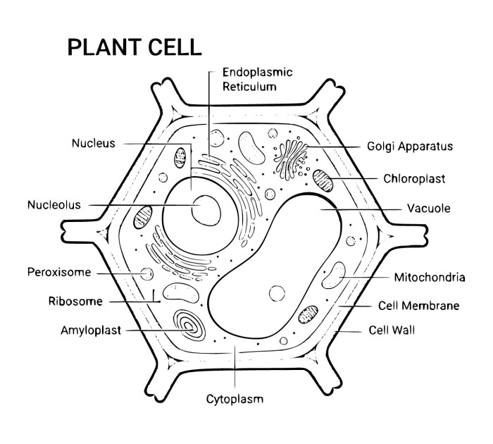
Plant and animal cells, while sharing some similarities, exhibit key differences that are crucial to understand. Plant cells possess a rigid cell wall, providing structural support and protection, a feature absent in animal cells. Furthermore, plant cells contain chloroplasts, the sites of photosynthesis, enabling them to produce their own food. This ability is not present in animal cells, which rely on consuming other organisms for energy.
Another significant difference lies in the presence of a large central vacuole in plant cells, responsible for storing water, nutrients, and waste products. This vacuole is significantly smaller or absent in animal cells. Finally, the shape of the cells themselves is often markedly different; plant cells tend to be more rigid and geometric due to the cell wall, whereas animal cells are typically more flexible and irregular in shape.
By highlighting these key features, the coloring pages become a valuable tool for comparing and contrasting these two essential cell types.
Designing Effective Coloring Pages
Creating engaging and informative coloring pages requires careful consideration of design elements to ensure they effectively communicate complex biological concepts like the structures of plant and animal cells. A well-designed coloring page should be visually appealing, easy to understand, and accurately represent the subject matter. The goal is to make learning fun and accessible.
Effective coloring pages utilize clear visuals, concise labels, and an appropriate level of detail suitable for the target audience. Overly complex designs can be overwhelming, while overly simplistic ones may lack educational value. The balance lies in creating a visually stimulating yet easy-to-follow representation of the cell’s components.
Plant Cell Coloring Page Design, Coloring pages plant and animal cells labeled
This coloring page will depict a plant cell, emphasizing its key organelles. The visual representation will utilize a simple, yet accurate, illustration of a plant cell. The design will incorporate a table to clearly organize the labels and their corresponding locations within the cell.
| Organelle | Description | Color Suggestion | Location in Cell |
|---|---|---|---|
| Cell Wall | Rigid outer layer providing support and protection. | Light Brown | Outermost layer, surrounding the cell membrane. |
| Chloroplasts | Sites of photosynthesis, containing chlorophyll. | Green | Scattered throughout the cytoplasm. |
| Vacuole | Large, central fluid-filled sac for storage and support. | Light Blue | Occupies a large portion of the cell’s interior. |
| Nucleus | Contains the cell’s genetic material (DNA). | Purple | Located near the center of the cell. |
| Cytoplasm | Gel-like substance filling the cell, containing organelles. | Light Yellow | Fills the space between the cell membrane and the organelles. |
Animal Cell Coloring Page Design
This coloring page will illustrate an animal cell, focusing on its major components. The design will prioritize clarity and accuracy, making it easy for users to identify and understand the function of each organelle. A bulleted list will provide concise descriptions of each organelle’s role within the cell.
The animal cell illustration will be simplified to avoid overwhelming detail, while still accurately representing the key organelles.
- Nucleus: Contains the cell’s genetic material (DNA) and controls cell activities.
- Cytoplasm: Gel-like substance filling the cell, containing organelles and providing a medium for cellular processes.
- Mitochondria: “Powerhouses” of the cell, responsible for energy production (ATP).
- Ribosomes: Sites of protein synthesis, crucial for building cellular components.
- Cell Membrane: Outer boundary of the cell, regulating the passage of substances in and out.
Comparison of Plant and Animal Cell Coloring Page Designs
The key difference in designing effective coloring pages for plant and animal cells lies in highlighting the unique organelles present in each. Plant cells require the inclusion of the cell wall, chloroplasts, and a large central vacuole, which are absent in animal cells. Animal cells, conversely, lack these structures but contain structures like centrioles (which can be optionally included for a more advanced coloring page) not found in plant cells.
Both designs should utilize clear, concise labels and visually distinct color schemes to differentiate the organelles, ensuring ease of understanding and memorization.
Coloring pages of plant and animal cells labeled offer a fantastic way to learn biology, but sometimes you need a break from the microscopic world! For a fun change of pace, check out these coloring pages animals Noah’s Ark , perfect for younger learners. Then, jump back to those plant and animal cell diagrams – you’ll appreciate the detail even more after a bit of creative animal fun.
Labeling Accuracy and Clarity: Coloring Pages Plant And Animal Cells Labeled

My dear students, the accuracy and clarity of labels on your coloring pages are paramount. Think of these labels as signposts guiding your understanding of the intricate world of plant and animal cells. Precise labeling transforms a simple coloring exercise into a powerful learning tool, solidifying your knowledge and fostering a deeper appreciation for cellular biology. A well-labeled diagram is not just a picture; it’s a roadmap to cellular comprehension.Clear and concise labels are essential for optimal learning because they directly link the visual representation of the cell components with their names.
Without accurate labels, the coloring page becomes a mere picture, failing to achieve its educational purpose. Ambiguous or inaccurate labels can lead to misconceptions and hinder the learning process. Remember, my young scholars, the goal is not just to color, but to learn. Let your labels be a testament to your understanding.
Font Styles and Sizes for Optimal Readability
Choosing the right font and size is crucial for ensuring that your labels are easily readable. Avoid overly decorative or stylized fonts; they can hinder comprehension. Sans-serif fonts, such as Arial or Calibri, are generally preferred for their clean lines and clear readability. Serif fonts, like Times New Roman, can also be used, but their more decorative nature might make them less suitable for smaller labels.
The font size should be large enough to be easily read without magnifying glasses, yet small enough to avoid overcrowding the illustration. A size between 8 and 12 points is generally suitable, depending on the overall size of the coloring page and the complexity of the illustration. Consider using bolding for main cell components to improve prominence. For example, “Nucleus” could be presented in bold 10-point Arial, clearly distinguishing it from other components labeled in regular 8-point Arial.
Strategic Label Placement for Clarity
The placement of labels is as important as their content. Poorly placed labels can clutter the illustration, making it difficult to understand. Strategically position labels to avoid obscuring important cellular structures. One effective method is to use leader lines—short lines connecting the label to the specific cell component—to guide the viewer’s eye. These lines should be thin and unobtrusive, preventing them from distracting from the overall illustration.
Avoid overlapping labels, and ensure sufficient spacing between them. Consider using a consistent color for all labels to maintain visual harmony. For instance, if you are labeling a chloroplast, you might place the label near it but slightly away, connecting them with a delicate, thin line. This prevents the label from covering the chloroplast itself while clearly identifying it.
If multiple labels are close together, consider using a numbered or lettered key to further clarify the relationship between labels and cell components.
Illustrative Examples and Descriptions
My dear students, let us delve into the captivating world of plant and animal cells, visualizing their intricate structures through detailed descriptions. Understanding these illustrations is key to grasping the fundamental differences and similarities between these vital units of life. We shall explore the nuances of their shapes, sizes, and the arrangement of their organelles, ensuring a clear and comprehensive understanding.
Plant Cell Illustration
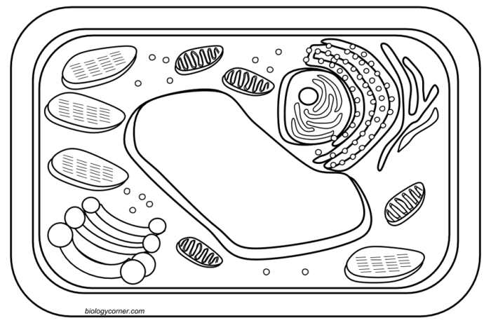
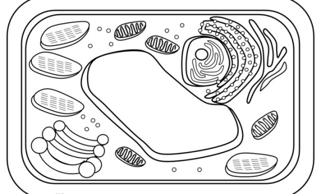
Imagine a plant cell, roughly rectangular in shape, significantly larger than its animal counterpart, perhaps measuring 10-100 micrometers in length. Its rigid structure is maintained by a sturdy cell wall, a cellulose-based outer layer, clearly visible in our illustration. Within this protective shell lies the cell membrane, a delicate, selectively permeable barrier controlling the entry and exit of substances.
The most striking feature, however, is the large central vacuole, occupying a substantial portion of the cell’s volume. This vacuole, depicted as a large, clear space, plays a crucial role in maintaining turgor pressure, giving the plant its firmness. The nucleus, often positioned centrally or slightly off-center, houses the genetic material. Scattered throughout the cytoplasm are numerous chloroplasts, the green energy factories responsible for photosynthesis.
These are depicted as oval-shaped organelles, their internal structure hinting at the complex processes within. Mitochondria, smaller and more numerous than chloroplasts, are also present, providing energy through cellular respiration. The endoplasmic reticulum, a network of interconnected membranes, is represented as a system of branching lines throughout the cytoplasm, reflecting its role in protein synthesis and transport. Finally, the Golgi apparatus, appearing as a stack of flattened sacs, modifies and packages proteins for secretion.
The presence of a large central vacuole and chloroplasts are defining characteristics that clearly distinguish plant cells from animal cells. These organelles are essential for plant growth, survival, and the process of photosynthesis, which sustains the entire plant kingdom.
Animal Cell Illustration
Now, let us turn our attention to the animal cell. Typically round or irregular in shape, an animal cell is generally smaller than a plant cell, ranging from 10-30 micrometers in diameter. Unlike its plant counterpart, it lacks a rigid cell wall. The cell membrane, however, remains the crucial boundary, regulating the passage of materials. The nucleus, often centrally located, is the command center, containing the cell’s genetic blueprint.
The cytoplasm is filled with various organelles. Mitochondria, essential for energy production, are numerous and scattered throughout. The endoplasmic reticulum, a complex network of membranes, is responsible for protein synthesis and transport. The Golgi apparatus, appearing as flattened sacs, modifies and packages proteins. Lysosomes, small, membrane-bound sacs, are involved in waste disposal and cellular recycling.
Ribosomes, tiny particles involved in protein synthesis, are scattered throughout the cytoplasm, either freely or attached to the endoplasmic reticulum. The centrosome, a region near the nucleus, plays a vital role in cell division.
The remarkable adaptability and diverse functions of animal cells are a testament to their intricate internal organization. The absence of a cell wall allows for greater flexibility in shape and movement, which is crucial for the various functions animal cells perform within the body.
Common Mistakes in Labeling Cell Diagrams
My young scholars, accuracy is paramount when labeling cell diagrams. Common mistakes include mislabeling organelles, incorrect positioning of organelles, and neglecting to label key structures altogether. For instance, confusing the Golgi apparatus with the endoplasmic reticulum, or misplacing the nucleus, are frequent errors. To avoid these pitfalls, it is crucial to consult reliable sources, study clear diagrams, and meticulously check your labels before finalizing your work.
Remember, precision is the hallmark of a true scholar. Understanding the functions of each organelle will help you accurately place and label them in the correct position within the cell. A well-labeled diagram reflects a deep understanding of cellular structure and function. Always double-check your work against trusted sources to ensure accuracy.
Educational Applications and Extensions

These coloring pages offer a vibrant and engaging pathway to understanding the intricate world of plant and animal cells. Their application extends far beyond simple coloring, serving as a powerful tool for reinforcing learning in various educational settings and fostering a deeper appreciation for biological structures. The interactive nature of the activity makes learning fun and memorable, promoting knowledge retention in a way that traditional methods may not achieve.These coloring pages can be seamlessly integrated into diverse learning environments, enriching the educational experience for students of all backgrounds.
The visual nature of the activity caters to different learning styles, making complex biological concepts more accessible and comprehensible. The detailed labeling further enhances understanding, allowing students to connect visual representations with terminology.
Classroom Integration
In a classroom setting, these coloring pages can be used as a pre-lesson activity to introduce the concepts of plant and animal cells, or as a post-lesson reinforcement exercise to solidify understanding. They can be incorporated into various lesson plans, used as individual assignments, group projects, or even as part of a larger unit on cell biology. Teachers can use them to assess student understanding by observing their labeling accuracy and comprehension of the different cellular components.
For instance, a teacher could assign students to color and label different organelles and then hold a class discussion comparing and contrasting plant and animal cells. Differentiated instruction can be easily implemented; students who grasp concepts quickly can research and add additional details, while those needing more support can focus on the basic labeling.
Homeschooling Applications
For homeschooling families, these coloring pages provide a flexible and engaging educational resource. They can be integrated into science lessons, used as a break from more traditional learning methods, or as part of a science-themed day. The self-directed nature of the activity allows children to work at their own pace, fostering independence and self-learning. Parents can use the coloring pages as an opportunity to engage in one-on-one learning, guiding their children through the labeling process and answering any questions they may have.
This personalized approach caters to individual learning needs and strengthens the parent-child bond.
Supplementary Activities
A series of supplementary activities can significantly enhance the learning experience derived from these coloring pages. These activities encourage deeper engagement with the material and promote a more holistic understanding of plant and animal cell structure and function.
- Quizzes: Simple quizzes, either written or oral, can assess student comprehension of the labeled structures and their functions. These can range from simple identification tasks to more complex questions about the roles of specific organelles.
- Research Projects: Students can delve deeper into the functions of specific organelles, researching their roles and importance within the cell. This could involve presentations, essays, or even creating models of the organelles.
- Comparative Analysis: Students can compare and contrast the structures and functions of plant and animal cells, highlighting the key differences and similarities between the two cell types. This activity fosters critical thinking and analytical skills.
Age Appropriateness
These coloring pages can benefit a wide range of age groups, adapting to different developmental stages and learning capabilities.Elementary school students (ages 6-10) can benefit from the visual learning and basic labeling, focusing on the major organelles and their general functions. The simplicity of the task helps build foundational knowledge in a fun and engaging way.Middle school students (ages 11-14) can engage with more complex labeling, exploring the specific functions of each organelle and comparing plant and animal cells.
They can also undertake more advanced research projects based on the coloring page activity.High school students (ages 15-18) can use the coloring pages as a review tool or as a foundation for more in-depth study of cell biology. They can utilize the pages to create more complex diagrams, including additional details and integrating their knowledge of cellular processes.

