Understanding Mycobacterium Tuberculosis Morphology

Mycobacterium tuberculosis drawing easy – Yo, Jogja peeps! Let’s dive into the world ofMycobacterium tuberculosis*, the bacteria behind tuberculosis (TB). Understanding its shape and structure is key to grasping how this sneaky bug causes disease. Think of it like understanding the blueprint of a villain’s lair before you can defeat them!
Basically,
-M. tuberculosis* is a rod-shaped bacteria, also known as a bacillus. Picture a tiny, slightly curved sausage. These bacilli are pretty small, typically measuring around 0.5 to 4 micrometers in length and about 0.2 to 0.5 micrometers in width. That’s seriously microscopic; you’d need a powerful microscope to even see them!
Mycobacterium Tuberculosis Key Structural Features
A simplified drawing of
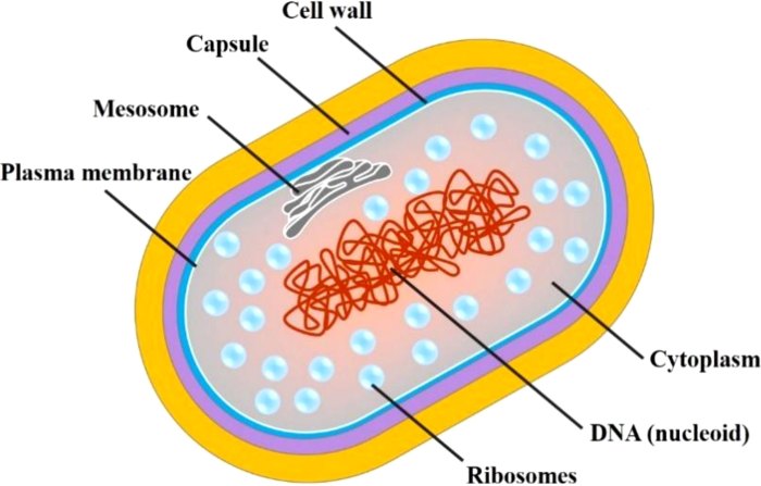
-M. tuberculosis* would highlight its crucial features. The most important part is its cell wall – a super thick and waxy outer layer made of mycolic acids. This waxy coat is what makes
-M. tuberculosis* acid-fast, meaning it resists staining with ordinary dyes.
It’s also what makes it so resilient and difficult to kill. Inside the cell wall lies the cytoplasm, containing the bacterium’s genetic material and other essential components. You’d also see a cell membrane just inside the cell wall.
Simplified Drawing Versus Microscopic Image
A simplified drawing focuses on the key structural features, emphasizing the bacillus shape, the thick cell wall, and the overall size. It’s a visual representation that helps us understand the basic anatomy. A microscopic image, however, reveals much more detail. You’d see finer structures within the cell, variations in shape and size among individual bacteria, and possibly even artifacts from the staining process.
The simplified drawing is like a schematic diagram, while the microscopic image is a detailed photograph.
Illustrative Line Drawing
Imagine a slightly curved line, about three times longer than it is wide. This represents the bacillus shape. Around this line, draw a thicker, slightly irregular outer layer representing the waxy cell wall. This outer layer should be noticeably thicker than the inner line representing the cell membrane. The space between the cell wall and membrane represents the cytoplasm.
This simple drawing effectively illustrates the key features of
-M. tuberculosis*: its rod shape, its prominent, thick cell wall, and the relatively smaller inner area representing the cell’s contents. Keep it simple; the detail is in the relative thicknesses of the layers, not in intricate inner structures.
Creating Educational Materials

Making learning about Mycobacterium tuberculosis fun and easy for Jogja’s young minds requires creative educational materials. We need to move beyond boring textbooks and engage students with visuals and interactive activities that stick. Think bright colours, relatable examples, and a touch of that unique Jogja vibe!
While depicting Mycobacterium tuberculosis requires a nuanced understanding of its cellular structure, simplifying the drawing for educational purposes is achievable. The challenge lies in accurately representing its key features, unlike the arguably simpler representations found in a resource like biomass easy diagram drawing , which focuses on a different level of biological complexity. Ultimately, effective Mycobacterium tuberculosis drawings prioritize clarity over intricate detail.
Here’s how we can create effective educational materials to help students understand the morphology of this tricky bacterium.
Labeled Diagram of Mycobacterium tuberculosis
A clear, labeled diagram is essential. Imagine a simple drawing of a rod-shaped bacterium, slightly curved. Label key features: the cell wall (emphasizing its thickness and waxy mycolic acid layer), the cytoplasm, and the nucleoid (representing the bacterial DNA). Use bright, contrasting colours to highlight these structures. For example, the cell wall could be a deep teal to represent its density, while the cytoplasm could be a lighter shade of green.
The nucleoid could be shown as a darker, more concentrated area of purple within the cytoplasm. Adding a scale bar would also help students understand the size of the bacterium (approximately 0.5-4 µm in length). A simple caption like “Mycobacterium tuberculosis: Note the thick, waxy cell wall” would provide context.
Worksheet with Blank Diagram for Completion
A worksheet incorporating a blank Artikel of the Mycobacterium tuberculosis bacterium encourages active learning. Provide students with a simple, unlabeled drawing of the rod-shaped bacterium. Then, include questions that guide them to label the key structures (cell wall, cytoplasm, nucleoid) based on what they’ve learned. You could also add questions about the bacterium’s size, shape, and unique characteristics, prompting them to think critically about what they’ve learned.
Perhaps include a section for them to draw the bacterium with its different structures colored according to their function. This hands-on approach helps reinforce their understanding.
Simplified Visual Representations
To simplify the complexity, we can use different visual approaches. One approach is to create an analogous representation, such as comparing the thick cell wall to a protective coat of armor. Another is to use a flowchart that illustrates the steps involved in the bacterium’s infection process. We can also use a 3D model made of clay or play-dough, showing the cell wall, cytoplasm, and nucleoid.
This tactile learning method can be particularly effective for visual and kinesthetic learners. Finally, a simple animation showing the bacterium’s movement could also help students visualize its behavior.
Caption for a Simple Drawing
A simple drawing showing a rod-shaped bacterium with a thick outer layer could be captioned as follows: “Mycobacterium tuberculosis: Notice the thick cell wall, a key feature making it resistant to many antibiotics.” This short caption highlights a crucial aspect of the bacterium’s biology, making it relevant to students’ understanding of tuberculosis treatment.
Advanced Drawing Techniques (Optional): Mycobacterium Tuberculosis Drawing Easy
Level up your Mycobacterium tuberculosis drawing skills! This section dives into some more advanced techniques to make your illustrations truly pop and accurately represent the complexity of this bacterium. We’ll explore shading, 3D representation, and showing its interaction with host cells. Get ready to impress your lecturers!
Mastering these techniques will allow you to create more realistic and informative diagrams, essential for understanding the nuances of this important pathogen. Think of it as adding those extra details that truly separate a good drawing from a great one – a drawing that really explains the science.
Bacterial Cell Wall Representation
The cell wall of Mycobacterium tuberculosis is unique, contributing to its pathogenicity. Accurately depicting this requires careful shading. You can use cross-hatching to show the dense, waxy layer of mycolic acids. Varying the density of the cross-hatching can illustrate variations in the cell wall thickness or different regions. For example, lighter shading could represent thinner areas, while darker, denser cross-hatching could show thicker regions.
Adding subtle gradients can also enhance the three-dimensional effect, creating a sense of depth and texture. Consider using a combination of pencil shading and stippling (small dots) for a more nuanced representation of the cell wall’s complexity.
Three-Dimensional Representation of the Bacterium
To create a three-dimensional effect, start by sketching the bacterium in its basic rod shape. Then, use shading and highlighting to create the illusion of volume. Remember, light sources affect how shadows are cast. A light source from above will create shadows beneath the bacterium, while a light source from the side will create shadows on the opposite side.
Use varying shades of gray to create depth and form. You can also add highlights to the areas where light would directly hit the bacterium, making it appear more three-dimensional. Think of how a curved surface reflects light differently from a flat one. The use of perspective can further enhance the 3D effect. Consider drawing the bacterium slightly angled to showcase its length and cylindrical shape more effectively.
Visual Cues for Bacterium-Host Cell Interaction, Mycobacterium tuberculosis drawing easy
Illustrating the interaction between M. tuberculosis and host cells requires careful planning. You can represent the phagocytosis process (where a host cell engulfs the bacterium) by drawing a macrophage (a type of immune cell) surrounding the bacterium. Use different colors and textures to distinguish the bacterium from the host cell. You can also depict the bacterium within a phagosome (a vesicle inside the macrophage) to illustrate its intracellular survival strategy.
Arrowheads can effectively indicate the direction of movement or interaction, for example, showing the bacterium entering the macrophage or the release of toxins. Using different colors and patterns for the macrophage’s cytoplasm and the phagosome can improve clarity and understanding.
Comparative Illustration of Bacteria
A comparative illustration is a great way to highlight the unique characteristics of M. tuberculosis. Consider placing M. tuberculosis alongside other bacteria like Escherichia coli (a rod-shaped bacterium) and Staphylococcus aureus (a spherical bacterium). Maintain consistent scale to accurately represent the relative sizes. Use clear labels to identify each bacterium. Highlight the differences in shape, size, and cell wall structure.
The differences in cell wall staining (acid-fast staining for M. tuberculosis versus Gram staining for others) could be subtly shown through color and texture variations in your illustration, although a direct representation of staining techniques is not necessary.
Common Queries
What are the best tools for drawing
-Mycobacterium tuberculosis*?
Pencils, pens, colored pencils, and digital drawing software are all suitable, depending on your skill level and preference.
How important is accuracy in a simplified drawing?
While simplification is key, maintaining the essential features (shape, cell wall) is crucial for accuracy and understanding.
Can I use color in my drawing?
Yes! Color can enhance understanding and make the drawing more engaging. Consider using colors to represent different parts of the bacterium or to highlight key features.
Where can I find more information on
-Mycobacterium tuberculosis*?
Reputable medical and scientific websites, textbooks, and educational resources offer detailed information on this bacterium.

